|
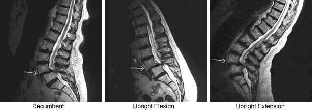
Recumbent only imaging can underestimate the extent of the pathology responsible for a patient's back pain by failing to visualize that pathology in the complete range of physiologic positions the spine normally occupies. In this case the recumbent scan showed an anterolisthesis at L4/5. The upright flexion scan showed an exaggeration of the L4/5 anterolisthesis. The upright extension scan showed a reduction of the anterolisthesis. In addition the dynamic Multi-Position UPRIGHT® MRI showed a fluctuating central canal stenosis that was most severe in UPRIGHT® flexion position. |