|
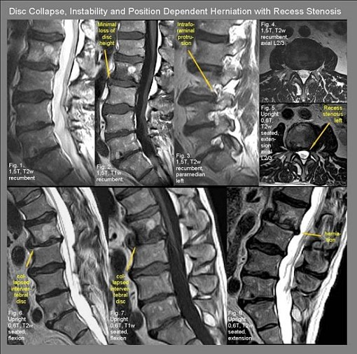
This 63-year-old man has been suffering from low back pain for years. Due to an increase in pain and new irradiation of his pain along the left L3 dermatome a recumbent MRI was performed. The recumbent MRI showed degenerative disc disease at L2/3, L4/5, and L5/S1 with Modic type changes indicating edema within the lower endplate of l2 (Fig.1, 2). A left intraforaminal disc protrusion was visible in the recumbent image at L3/4 and was thought to be responsible for the patient's radicular symptoms (Fig 3). However when the patient's symptoms continued to deteriorate his physician referred him for an UPRIGHT® Multi-Position MRI evaluation that showed a striking loss of disc height at L2/3 that was not visible on the recumbent MRI (Fig.6, 7) accompanied by pronounced posterior and anterior herniations of disc material (Fig.6, 7, 8) kyphosis (Fig.6), postero-listhesis at L2/3 (Fig.8) and canal stenosis (fig.5) that was either not visualized at all by recumbent MRI or to an extent that significantly underestimated the pathology relative to its magnitude in the UPRIGHT® position. (E.g. recumbent disc herniations seen at L2/3 compared to those seen in the UPRIGHT® positions) |